Left Hip Muscles Anatomy : Tendinitis And Bursitis Treatment Cincinnati Tendinitis Dayton Oh : 1 hip anatomy, function and common problems.
byLeanne Moore•
0
Left Hip Muscles Anatomy : Tendinitis And Bursitis Treatment Cincinnati Tendinitis Dayton Oh : 1 hip anatomy, function and common problems.. It is a flat, triangular muscle on the anterior wall of the pelvis. If left unstretched, shortened hip flexors affect the position of the pelvis, which in turn affects the position and movement of the lower back. If you know all the hip flexor names and bones they attach to, that's an awesome accomplishment! There are three layers of gluteal muscles on the posterior hips, just like there are three layers of muscles in the abdominal trunk. In order to isolate the abdominals, you need to minimize the involvement of the hip flexors and maximize the contraction of the abdominals.
Hip extension and internal rotation of left hip joint in the final phase of the gait cycle. There are a lot of muscles of the hip and thigh. Groin, inguinal region and the anterior. Advanced hip flexor muscle anatomy. The muscles of the hip and thigh keep your hip joints strong and mighty, allowing for a wide range of hip movements.
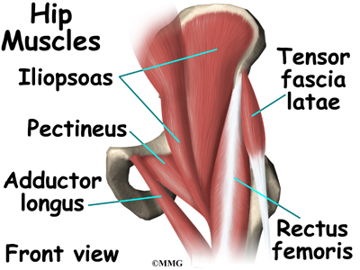
Hip Anatomy Eorthopod Com from eorthopod.com The anterior boundary of the hip adductors is set by if left unchecked, this can lead to chronic knee pain from it band syndrome or acute yet severe injuries such as knee ligament tears (e.g. If you know all the hip flexor names and bones they attach to, that's an awesome accomplishment! Rectus femoris forms the middle portion of the quadriceps. Learning the anatomy of your hip will better enable you to pinpoint your pain and work with your doctor to keep it from limiting your life. The hip's essential muscles are the sartorius, rectus femoris, gluteus minimus and medius, iliopsoas, adductors, and hamstrings. The different anatomical areas of the gluteal region: Most modern anatomists define 17 of these muscles, although some additional muscles may sometimes be considered. I pulled some muscles on left hip hiking.
Pelvis and acetabulum, with muscle attachment sites.
Understanding the anatomy of the lower body, particularly the muscle locations and their functions, will help you to get the most from the exercises and programs presented on this website. These muscles constitute the anatomical classification known as the medial compartment of the thigh. Learn their anatomy efficiently and easily using kenhub's muscle anatomy and reference charts! Muscle movements, types, and names. The muscles of the neck can be divided into groups according to their location. Left leg, lateral (left) and posterior (right) views. There are a lot of muscles of the hip and thigh. The muscles of the pelvis, hip and buttock anatomical chart shows how each muscle in this area of the body works with the others, and the various minor systems within the major ones. It originates at the anterior inferior iliac spine and just above the acetabulum of the hip bone. If left unstretched, shortened hip flexors affect the position of the pelvis, which in turn affects the position and movement of the lower back. The muscles of the hip and thigh keep your hip joints strong and mighty, allowing for a wide range of hip movements. The cavity of the acetabulum the external obturator muscle is short external rotator muscle of hip joint. Microscopic anatomy of skeletal muscle.
Microscopic anatomy of skeletal muscle. If left unstretched, shortened hip flexors affect the position of the pelvis, which in turn affects the position and movement of the lower back. Muscles of the hips and thighs | human anatomy and. 1 hip anatomy, function and common problems. Anatomy of the muscular system.
Muscles Of The Hips And Thighs Human Anatomy And Physiology Lab Bsb 141 from s3-us-west-2.amazonaws.com Rectus femoris forms the middle portion of the quadriceps. The hip joint is the articulation of the pelvis with the femur, which connects the axial skeleton with the lower extremity. Major lower body muscle groups include leg and hip muscles, largest muscle groups in your body. The muscular system is responsible for the movement of the human body. Understanding the anatomy of the lower body, particularly the muscle locations and their functions, will help you to get the most from the exercises and programs presented on this website. Anterior muscles extend your legs and flex your thighs. It originates at the anterior inferior iliac spine and just above the acetabulum of the hip bone. Advanced hip flexor muscle anatomy.
The muscles of the pelvis, hip and buttock anatomical chart shows how each muscle in this area of the body works with the others, and the various minor systems within the major ones.
The muscles of the neck can be divided into groups according to their location. Muscles that act on the lower limb cause movement at the hip, knee and foot joints. Anatomy of the muscular system. Learning the anatomy of your hip will better enable you to pinpoint your pain and work with your doctor to keep it from limiting your life. If you know all the hip flexor names and bones they attach to, that's an awesome accomplishment! There are three layers of gluteal muscles on the posterior hips, just like there are three layers of muscles in the abdominal trunk. Attached to the bones of the skeletal system are about 700 named. Muscle movements, types, and names. Anatomy of a human body we study anatomy. Leave a reply cancel reply. Most modern anatomists define 17 of these muscles, although some additional muscles may sometimes be considered. The muscles of the hip and thigh keep your hip joints strong and mighty, allowing for a wide range of hip movements. This anatomical atlas was especially designed for a specific public (radiologists, surgeons, rheumatologists and physicians specializing in musculoskeletal imaging).
I pulled some muscles on left hip hiking. The hip joint is the articulation of the pelvis with the femur, which connects the axial skeleton with the lower extremity. The muscles of the neck can be divided into groups according to their location. The muscles of the pelvis, hip and buttock anatomical chart shows how each muscle in this area of the body works with the others, and the various minor systems within the major ones. Muscles of the hips and thighs | human anatomy and.
Hip Muscle Strains Info Florida Orthopaedic Institute from viewmedica.com The main functions of the neck muscles are to permit movements of the neck or head and to provide structural support of the head. Now that you watched the video, you. If left unstretched, shortened hip flexors affect the position of the pelvis, which in turn affects the position and movement of the lower back. The hip's essential muscles are the sartorius, rectus femoris, gluteus minimus and medius, iliopsoas, adductors, and hamstrings. Included within the chart are gorgeous illustrations of the pelvic diaphragm, sphincter muscles, gluteus maximus. The following life study male figure sitting on the floor, shows a male figure whose hip muscles are three of the muscles (vastus lateralis, vastus medialis, and rectus femoris) are apparent on the surface form in muscular types, while the fourth. In order to isolate the abdominals, you need to minimize the involvement of the hip flexors and maximize the contraction of the abdominals. This 20 x 26 (51 x 66 cm) wall poster shows location of various joints and provides anterior and posterior views of the left shoulder, right hip, right knee and left elbow.
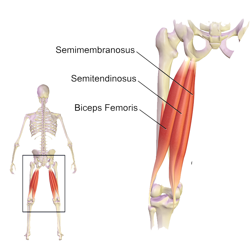
These muscles are responsible for hip joint extension (backward movement).
Included within the chart are gorgeous illustrations of the pelvic diaphragm, sphincter muscles, gluteus maximus. Learn their anatomy efficiently and easily using kenhub's muscle anatomy and reference charts! Your email address will not be published. Major lower body muscle groups include leg and hip muscles, largest muscle groups in your body. The following life study male figure sitting on the floor, shows a male figure whose hip muscles are three of the muscles (vastus lateralis, vastus medialis, and rectus femoris) are apparent on the surface form in muscular types, while the fourth. 1 hip anatomy, function and common problems. The gluteus medius muscle helps abducts the thigh along with the gluteus maximus, but can rotate the thigh inward where the gluteus maximus rotates the thigh outward. for detailed anatomy of pelvic bones, read anatomy of hip bone. Learn about hip muscles human anatomy with free interactive flashcards. Groin, inguinal region and the anterior. Now that you watched the video, you. This webpage presents the anatomical structures found on hip mri. One example of an ab exercise that actually focuses.